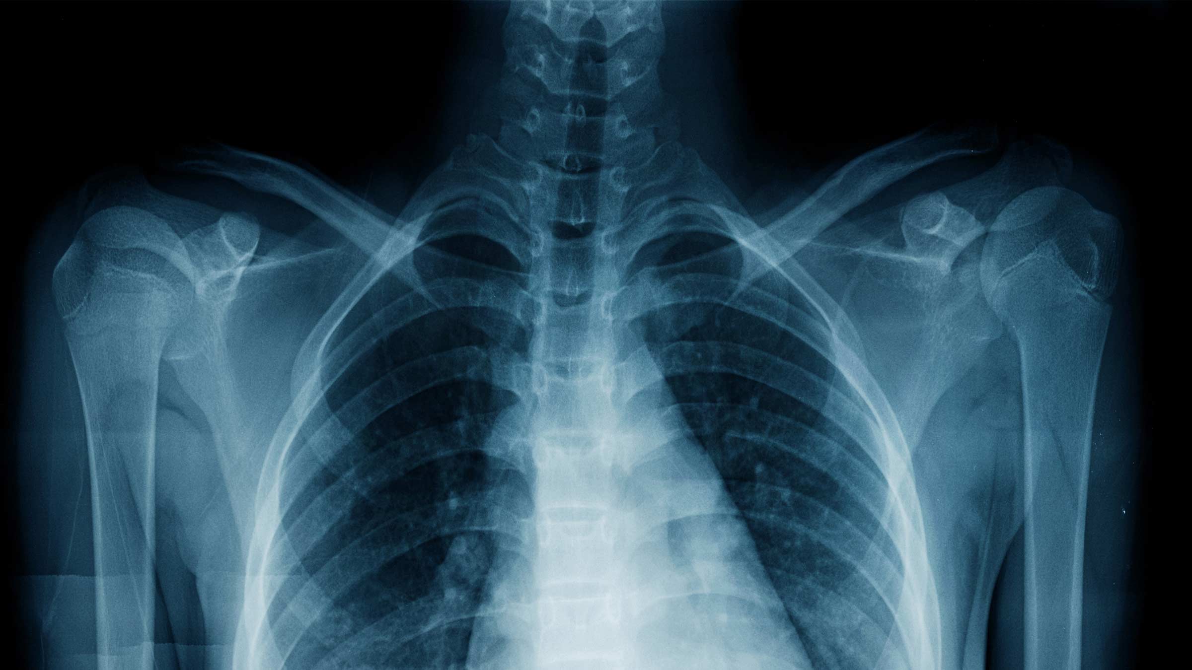
Unveiling Male Anatomy: The Science Of Penile Imaging
The human body is a marvel of biological engineering, and for centuries, our understanding of its inner workings was limited to what could be observed externally or through invasive procedures. However, with the advent of advanced medical imaging technologies, we've gained unprecedented access to the intricate structures and dynamic processes within. Among these fascinating areas of study is the male anatomy, particularly the penis, and how it can be visualized for both diagnostic purposes and scientific inquiry. While the phrase "xray of cock" might conjure various images, its true significance lies in its capacity to reveal crucial medical insights.
This article delves into the sophisticated world of penile imaging, exploring the various techniques used, their applications in diagnosing conditions, and their role in advancing our understanding of male sexual health. We'll navigate the complexities of visualizing this vital organ, distinguishing between scientific exploration and common misconceptions, and highlighting why accurate medical assessment is paramount for well-being.
Table of Contents
- Unveiling the Invisible: The Science of Penile Imaging
- A Glimpse into Anatomy: What "X-ray of Cock" Truly Means in Medicine
- Advanced Imaging Techniques for Male Reproductive Health
- Diagnosing Rare Conditions: The Case of Penile Ossification
- Imaging in Sexual Health Research: Understanding Intimacy
- The Ethics and Misconceptions of "X-ray of Cock" in Public Discourse
- The Future of Penile Imaging: Innovations and New Discoveries
- Prioritizing Health: When to Seek Professional Medical Advice
Unveiling the Invisible: The Science of Penile Imaging
The human body, in all its complexity, often holds secrets within that are not visible to the naked eye. Medical imaging has revolutionized our ability to peer inside, offering invaluable diagnostic tools and insights into physiological processes. When we talk about "xray of cock," we're essentially referring to the broader field of imaging the male reproductive organ, which encompasses a range of sophisticated technologies far beyond just traditional X-rays. This scientific endeavor is crucial for understanding normal anatomy, diagnosing pathologies, and even exploring the dynamics of sexual function.
Beyond the Naked Eye: Why Imaging Matters
Why is it so important to see inside? For one, many conditions affecting the penis, such as Peyronie's disease, erectile dysfunction, or even trauma, involve internal structures like the erectile tissues, blood vessels, and nerves. External examination alone often isn't enough to pinpoint the exact cause or extent of a problem. Imaging provides a non-invasive or minimally invasive way to visualize these internal components, allowing healthcare professionals to make accurate diagnoses and formulate effective treatment plans. Without these tools, many conditions would remain undiagnosed or poorly understood, leading to suboptimal care. The ability to obtain a detailed "xray of cock," in its broader sense, empowers medical practitioners to provide precise and personalized care.
A Glimpse into Anatomy: What "X-ray of Cock" Truly Means in Medicine
While the term "xray of cock" might be casually used, in a medical context, it refers to the application of various imaging modalities to visualize the penis. Traditional X-rays, which use ionizing radiation to create images of bones and dense structures, are generally not the primary choice for soft tissue organs like the penis. This is because the penis is predominantly composed of soft tissues (erectile tissue, smooth muscle, blood vessels, nerves), which do not show up well on standard X-ray films unless they are calcified or contain foreign bodies. However, modified X-ray techniques, such as cavernosography (where a contrast dye is injected into the erectile tissue), can be used to assess blood flow issues or structural abnormalities. The term, therefore, serves as a general descriptor for visualizing the male anatomy internally, though more advanced techniques are often preferred for their superior soft tissue resolution.
Advanced Imaging Techniques for Male Reproductive Health
Beyond basic X-rays, several sophisticated imaging techniques offer detailed views of the male anatomy, each with its unique strengths and applications. These methods provide critical information for diagnosing a wide array of conditions affecting the penis and male reproductive system.
MRI: Revealing Soft Tissues and Dynamic Processes
Magnetic Resonance Imaging (MRI) stands out as a powerful tool for visualizing soft tissues with exceptional clarity. Unlike X-rays, MRI uses strong magnetic fields and radio waves to generate detailed images, making it ideal for examining the complex structures of the penis, including the corpora cavernosa (erectile bodies), corpus spongiosum, and urethra. MRI is particularly valuable for diagnosing conditions like Peyronie's disease (fibrous plaques that cause penile curvature), tumors, and even for assessing the extent of trauma. The precision offered by an MRI can often negate the need for more invasive diagnostic procedures. Notably, as mentioned in scientific literature, "Magnetic resonance imaging was used to study the female sexual response and the male and female genitals during coitus," demonstrating its unique capability to capture dynamic physiological processes in real-time, offering unparalleled insights into sexual function and interaction.
Ultrasound and CT Scans: Complementary Views
Ultrasound, or sonography, is another widely used and non-invasive imaging technique. It employs high-frequency sound waves to create real-time images of internal structures. For the penis, ultrasound is excellent for evaluating blood flow (Doppler ultrasound), diagnosing erectile dysfunction, identifying plaques in Peyronie's disease, and detecting cysts or masses. Its real-time nature allows for dynamic studies, such as assessing blood flow changes during an induced erection. Computed Tomography (CT) scans, while also using X-rays, provide cross-sectional images that are more detailed than conventional X-rays. CT scans are less commonly used for primary penile imaging due to radiation exposure and less soft tissue contrast compared to MRI, but they can be useful in cases of severe trauma, suspected fractures, or when assessing the spread of cancer to surrounding bone structures.
Diagnosing Rare Conditions: The Case of Penile Ossification
Medical imaging plays a crucial role in identifying and understanding rare conditions that might otherwise go unnoticed or be misdiagnosed. One such intriguing case is penile ossification, a rare condition where bone forms abnormally within the soft tissues of the penis. This phenomenon, often referred to as "penile bone" or "os penis" in animals, is highly unusual in humans. The provided data highlights a specific instance: "A man was diagnosed with penile ossification, a rare condition in which bone forms inside the penis." Such a diagnosis typically relies heavily on imaging techniques like X-rays or CT scans, which are excellent at visualizing calcified structures. Understanding the presence and extent of such ossification is vital for managing symptoms, which can include pain, erectile dysfunction, and curvature, and for determining the appropriate course of treatment, which might involve surgical removal of the calcified tissue. This demonstrates the critical diagnostic power that an "xray of cock" (in the sense of a bone-focused image) can possess.
Imaging in Sexual Health Research: Understanding Intimacy
Beyond diagnosing pathologies, medical imaging has opened new frontiers in sexual health research, allowing scientists to study the physiological aspects of sexual response and interaction with unprecedented detail. The ability to visualize internal anatomy during coitus or other sexual activities provides invaluable data that was previously unobtainable. As noted in the provided data, "Thirteen experiments were performed with eight couples and three single women" using MRI to study sexual response. This type of research, while ethically complex and requiring careful consideration, contributes significantly to our understanding of human sexuality, reproductive health, and potential treatments for sexual dysfunctions. It allows researchers to observe changes in blood flow, muscle contraction, and anatomical relationships in real-time, offering insights into the mechanics of sexual function that no other method can provide. This scientific application of "xray of cock" (in the sense of internal visualization during sexual activity) is purely for academic and medical advancement.
The Ethics and Misconceptions of "X-ray of Cock" in Public Discourse
The phrase "xray of cock" often circulates in public discourse, sometimes with sensational or misleading connotations. It's important to distinguish between legitimate medical imaging and the portrayal of such concepts in non-scientific or even explicit contexts. The public's fascination with seeing "inside" has led to the proliferation of various forms of media, some of which purport to show "xray dick inside pussy" or "xray deepthroat oral dick throat free videos." These often employ visual effects or highly stylized content rather than genuine medical imaging, and they exist primarily for entertainment purposes. It is crucial to understand that reputable medical imaging is conducted under strict ethical guidelines, with patient consent, and for specific diagnostic or research objectives. The purpose is always to improve health outcomes or advance scientific knowledge, not for gratuitous display. Misconceptions can arise when the term "xray of cock" is encountered in contexts far removed from its medical utility, leading to confusion about what these technologies actually reveal and for what purpose. Trustworthy information about health and anatomy should always be sought from qualified medical professionals and peer-reviewed scientific sources, not from entertainment platforms.
The Future of Penile Imaging: Innovations and New Discoveries
The field of medical imaging is constantly evolving, and penile imaging is no exception. Advances in technology promise even more detailed, faster, and less invasive ways to visualize the male anatomy. Researchers are exploring new frontiers such as functional MRI (fMRI) to map brain activity related to sexual response, advanced ultrasound techniques for even finer resolution of microvasculature, and the integration of artificial intelligence (AI) for automated image analysis and diagnosis. AI could potentially assist in identifying subtle abnormalities, predicting treatment responses, and even personalizing imaging protocols for individual patients. The development of even more precise and patient-friendly imaging modalities will continue to enhance our understanding of male reproductive health, leading to earlier diagnoses, more effective treatments, and ultimately, improved quality of life for men facing urological or sexual health challenges. The ongoing quest to refine the "xray of cock" in its medical application holds immense promise for the future of healthcare.
Prioritizing Health: When to Seek Professional Medical Advice
Understanding the capabilities of penile imaging is valuable, but it's even more important to know when to seek professional medical advice. If you experience any symptoms related to your penile health, such as pain, swelling, lumps, curvature, changes in erectile function, or any other concerns, it is crucial to consult a qualified healthcare professional. Self-diagnosis based on anecdotal information or misleading content can be dangerous and delay proper treatment. A urologist or a general practitioner can assess your symptoms, recommend appropriate diagnostic tests (which may include various forms of "xray of cock" or other imaging), and provide an accurate diagnosis and treatment plan. Remember, your health is paramount, and reliable medical information and care should always come from trusted, certified sources.
Conclusion
The phrase "xray of cock," when viewed through a scientific and medical lens, unlocks a fascinating realm of anatomical understanding and diagnostic power. Far from being a mere curiosity, the various imaging techniques applied to the male anatomy – including advanced MRI, ultrasound, and specialized X-ray procedures – are indispensable tools in modern medicine. They enable healthcare professionals to diagnose rare conditions like penile ossification, assess common issues like erectile dysfunction, and contribute to groundbreaking research on human sexual response. By providing unparalleled insights into the body's internal workings, these technologies empower us to maintain and improve male reproductive health.
It is essential to distinguish between the legitimate, ethically-driven applications of medical imaging and any sensationalized or misleading portrayals. For accurate information and professional guidance regarding male health, always consult qualified medical experts and reputable scientific resources. Your well-being depends on informed decisions and trusted care. We encourage you to prioritize your health and seek professional advice for any concerns you may have, ensuring that the incredible capabilities of medical imaging are utilized for your benefit.
- Is Judge Lauren Lake Married
- Iran Is Safe
- Ann Stringfield
- Is Ice Spice Dating Anyone
- Stanley Marketplace
/studio-shot-of-chest-x-ray-136595862-595bdb493df78c4eb6a38dce-5c59d3ecc9e77c000132accf.jpg)
X Ray Definition and Properties (X Radiation)

X-ray and what to expect | Ohio State Health & Discovery

X-Rays: MedlinePlus